Botany Blog Plants of the Northeastern U.S.

February 28, 2010

Dark Septate Endophytes

Filed under: Mycorrhizae — admin @ 02:49

Dark septate endophytes (DSE) are a heterogenous group of conidial or sterile fungi (thought to be ascomycetes) with darkly-pigmented, septate hyphae that commonly colonize plant roots. Most DSE also produce intracellular structures called microsclerotia. It has been suggested that these microsclerotia may serve as dispersal structures (Currah et al., 1993), although the actual purpose appears to be unknown. Perhaps they serve a storage function for the fungi as do the vesicles of AM fungi?

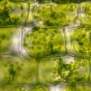
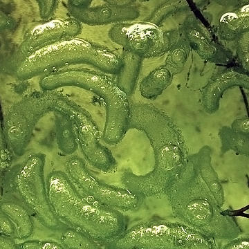
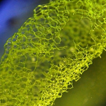
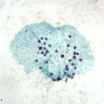
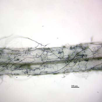
The following images show septate hyphae of a DSE growing in and around a root of Eurybia divaricata. The first image shows net-like strands formed by the hyphae of a DSE:

DSE covering a root of White Wood Aster

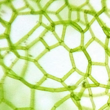
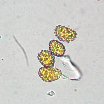
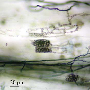
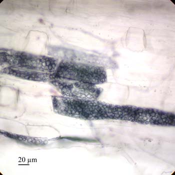
 The next image depicts several hyphae ending in microsclerotia, which appear as grape-like clusters.

Dark septate hyphae and miscrosclerotia

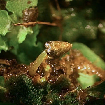
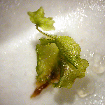
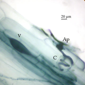
Another  morphotype can be seen with a root of Maianthemum racemosum in the following image:

Microsclerotia in a root of False Solomon’s Seal

The taxonomy of this group is poorly understood and little is known of the role that DSE play in natural ecosystems (see review by Jumpponen and Trappe 1998). DSE are difficult to identify from field samples so many may not yet be known. A few of the species identified to date (mostly from bioassays) include Chloridium paucisporum, Leptodontidium orchidicola, Phialocephala dimorphosphora, Phialocephala fortinii, and Phialophora finlandia. Of these, Phialocephala fortinii appears to be the best studied. Unlike AM fungi, the DSE that have been studied to date do not require a plant host and many can be grown as pure cultures.

Available evidence suggests that DSE range from strongly pathogenic to non-pathogenic to mycorrhizal for plants. Some strains of DSE may be involved in host plant nutrient acquisition and it has been proposed that this may be a mutualistic, mycorrhiza-like relationship (Jumpponen and Trappe 1998). These relationships do not appear to be host specific (Jumpponen and Trappe 1998), with some strains forming ectendomycorrhizas but also associating with other hosts in ways not completely understood (Jumpponen 2001).

Although their mycorrhizal status is uncertain (Jumpponen and Trappe 1998), DSE are commonly associated with the roots of herbaceous and woody plants from alpine, boreal and northern temperate ecosystems (Haselwandter and Read 1980, Holdenrieder and Sieber 1992; O’Dell et al. 1993; Ahlich and Sieber 1996). To date, carbohydrate flow from a host plant to a DSE has not yet been demonstrated as it has for AMF (Jumpponen 2001). Despite their ubiquity in terrestrial ecosystems, the role of DSE in plant nutrient acquisition and plant community assemblage remains largely unknown. Given the ubiquity of DSE in many terrestrial ecosystems and their possible mycorrhiza-like relationships with plants, studying this group of fungi could increase our understanding of plant community assemblage.

Literature Cited:

Ahlich, K. and T.N. Sieber. 1996. The profusion of dark septate endophytic fungi in non-ectomycorrhizal fine roots of forest trees and shrubs. New Phytologist 132:259-270.

Currah, R.S., Tsuneda, A., and Murakami, S. 1993. Morphology and ecology of Phialocephala fortinii in roots of Rhododendron brachycarpum. Canadian Journal of Botany 71:1639-1644.

Haselwandter, K. and D.J. Read. 1980. Fungal associations of roots of dominant and sub-dominant plants in high-alpine vegetation systems with special reference to mycorrhiza. Oecologia 4:557-62.

Holdenrieder, O. and T.N. Sieber. 1992. Fungal associations of serially washed healthy non-mycorrhizal roots of Picea abies. Mycological Research 96:151-156.

Jumpponen, A. and J.M. Trappe. 1998. Dark septate endophytes: a review of facultative biotrophic root-colonizing fungi. New Phytologist 140:295-310.

Jumpponon, A. 2001. Dark septate endophytes – are they mycorrhizal? Mycorrhiza 11:207-211.

O’Dell, T.E., H.B. Massicotte and J.M. Trappe. 1993. Root colonization of Lupinus latifolius Agardh. and Pinus contorta Dougl. By Phialocephala fortinii Wang & Wilcox. New Phytologist 124:93-100.

February 24, 2010

Plasmolysis

Filed under: Uncategorized — admin @ 02:04

Plant cells rely on internal hydrostatic pressure to maintain their shape. This pressure is maintained by a relatively high solute concentration inside the vacuole, which results in the absorption of water from outside the cell via osmosis (Raven et al., 2007). As the vacuole swells with water, the protoplast increases in size and the plasma membrane pushes up against the inside of the cell wall, resulting in turgor pressure. The turgor pressure is counteracted by the call wall and these opposing forces help maintain the shape of the cell and ultimately support herbaceous plant tissues.

The image below show cells in a leaf of Elodea canadensis that was placed in a solution of distilled water. The small green structures are the chloroplasts inside the cells and notice that they are distributed near the cell wall.

 Normal Plant Cell

Elodea cell in a solution of distilled water

If a plant does not receive sufficient water or is placed in environment that is hypertonic (one that has a higher solution concentration than the plant cells, e.g. a salty environment), than the cells will lose water and the plants will wilt. This is because water will be drawn out of the vacuole through osmosis, the protoplast will shrink, and the plasma membrane will actually pull away from the cell wall (plasmolysis), resulting in a loss of turgor pressure.

The following image shows an Elodea leaf that was placed in a 20% sucrose (sugar) solution. Note how the inside of the cell is shrinking and the gap between the plasma membrane and the cell wall (the cell wall does not shrink because it is somewhat rigid due to the presence of cellulose microfibrils).

Plasmolysed plant cell

 Elodea cell in a solution of 20% sucrose

Literature Cited

Raven, P.H., Evert, R.F., and S.E. Eichhorn. 2007. Biology of Plants, 7th ed. Worth Publishers, Inc., NY.

February 8, 2010

Eastern Comma

Filed under: Plant-Insect Interactions — admin @ 22:25

Spring semester is in full swing so it has been difficult to find time to write of late. I shot this image of an Eastern Comma (Polygonia comma) last summer as it was resting on a bench along the boardwalk that leads into a poor fen here in central NY.

Eastern Comma

This butterfly occurs throughout most of the eastern half of the United States and has two broods a year. The larvae will feed on a variety of host plants including False Nettle (Boehmeria cylindrica), Wood Nettle (Laportea canadensis), American Elm (Ulmus americana) and Stinging Nettle (Urtica dioica). Adults feed on the sap of trees (Layberry et al. 1998).

Literature cited:

Layberry, R.A., Hall, P.W., and Lafontaine, J.D. 1998. The Butterflies of Canada. University of Toronto Press.

January 17, 2010

Passiflora x belotii

Filed under: Plant-Insect Interactions — admin @ 02:31

Passiflora x belotii is the name given to the hybrid of P. alata and P. caerulea. It is a sterile hybrid that will not produce fruit but is a prolific bloomer when given plenty of light. The flower shown here was taken from one of my plants that was erroneously labeled P. incarnata at the nursery where I bought it.

Passiflora x belotii

There are over 450 recognized species in the genus Passiflora (Vanderplank 1996). Most are vines, but a small number can grow as trees (e.g. P. lindeniana). Most people recognize passionflowers as having purple and white flowers, but there are species that produce white, pink, yellow, red and even orange flowers. The flowers of the hybrid shown here are very fragrant with a scent similar to that of the fruit of P. edulis. The foliage of passionflowers is toxic primarily due to the presence of cyanogenic glycosides. When a leaf is chewed by an insect, these glycosides interact with ezymes in the leaf (much like when a glow stick is “broken” allowing the chemical components to mix) and release hydrogen cyanide as a by-product. A group of tropical and subtropical butterflies (subfamily Heliconiinae) have co-evolved with plants in the genus Passiflora and have developed the ability to somehow metabolize these compounds and use them as a defense against predators (Engler et al. 2000).  One of the better known species is the red postman butterfly, Heliconius erato, which is often displayed in butterfly houses at botanical gardens and nature centers.

Heliconius erato

This image was taken at Hershey Gardens in their butterfly house. It is worth noting that the garden does not breed or raise their butterflies; they purchase pupae from places in the tropics that rear caterpillars, some of which are sold while others are released back into the wild to aid conservation efforts in areas where suitable habitat is diminishing.

Engler, H.S., Spencer, K.C. and Gilbert, L.E. 2000. Insect metabolism: Preventing cyanide release from leaves. Nature, 406, 144-145.

Vanderplank, J. 1996. Passion Flowers, 2nd ed. The MIT Press. Cambridge, MA.

January 1, 2010

Water Net (Hydrodictyon reticulatum)

Filed under: Algae — admin @ 16:00

It would seem that the subject of my posts keep getting smaller and smaller. These images are of a common green algae (phylum Chlorophyta) commonly known as Water Net. It gets its name from the pentagonal or hexagonal branching pattern of the filaments making up the body (thallus) of the algae. These specimens came in on a sample of Chara, another green algae that is considered to be the closest living relative to plants. I took a sample of the Chara out the other day to photograph and left the sample in a dish of water under some lights. After about a week these little green ‘bubbles’ started appearing and have continued to get progressively larger.

Hydrodictyon reticulatum

I was really curious what this stuff was but couldn’t really find anything that fit the description. I was already aware that water net was growing in the tank where I am keeping the Chara, but the filaments on these were not visible to the naked eye. It wasn’t until I got out a hand lens that they became apparent.

hydrodictyon2.jpg

Apparently they can form these somewhat spherical colonies that just keep expanding as the thallus divides. I got one good closeup showing how each filament is connected to two other filaments, which creates the unique branching pattern and allows the colony to take on a 3-dimensional form.

hydrodictyon4.jpg

Algae in the genus Hydrodictyon are isogamous (gametes all alike) and the cells are coenocytic (multinucleate). Dictyo is Greek for net-like, so the genus means the same as the common name.

December 23, 2009

Scented Liverwort

Filed under: North American Native Plants,Seedless Plants — admin @ 05:46

I recently updated the images on the page for Conocephalum conicum (Scented Liverwort), primarily to add an image of the sporophyte. They look like really small mushrooms, and the dark, round spots at the base of the ‘cap’ are the sporangia. Meiosis takes place in the sporangia and they eventually release spores.

null

December 18, 2009

Venus flytrap

Here are some of the “traps” of a venus flytrap, each consisting of a bi-lobed leaf at the end of a flattened petiole. They are active traps, meaning that they are capable of closing quickly to ensnare a prey item. Each lobe of the trap is beset with three large trichomes (the trigger hairs), readily visible in the image below. If any two of the hairs are touched, or if one of them is touched twice, this will cause a rapid pressure change within the cells and trigger the trap to close. The long segments along the edge of each lobe may look like sharp teeth but they are quite soft to the touch; they serve to prevent the escape of the prey, not to impale it.

Venus Flytrap

Venus flytrap (Dionaea muscipula) is a very popular plant, mostly owing to its ability to actively capture and digest insects. It has become widely available now due to the ease by which it can be propagated using tissue culture. Unfortunately many who attempt to grow this plant fail due to a lack of understanding of its cultural requirements. Understanding how to successfully grow this plant begins with understanding where it comes from and why it captures bugs in the first place.

Venus flytrap is the only species in its genus. It is in the family Droseraceae, which includes another group of carnivorous plants known as sundews (Drosera spp., e.g. Drosera rotundifolia). Unlike sundews, the venus flytrap has a much more limited natural range, originally found only in a small area of southern North Carolina and northern South Carolina. The natural habitat of this plant is open, acidic wetlands on peaty or sandy soils with low nitrogen availability. The capture and digestion of insects is an adaptation to these nitrogen-poor environments, allowing the plant to break down proteins and absorb the nitrogen for use in metabolic functions.

One mistake people make in trying to grow this plant is not providing it with sufficient humidity. Dry air causes the leaves and the traps to dry out and turn black. A glass or plastic cover satisfies this requirement; a small gap in the cover should exist to allow for some air circulation. Watering can be accomplished by standing the pots in shallow water. Distilled water is best, as hard water or water containing various salts (this would include softened water) will eventually kill the plant. While the plants can stand being in soil that is completely saturated for short periods, it is best to avoid this.

The question I am most often asked is if it is necessary to feed them. Not really, at least not as often as some would think. It is also best to feed the plants insects and not hamburger, although lean meats will work. I prefer to either set plants outside for a time in the summer to allow them to capture insects naturally or give a light dose of fertilizer to the leaves.

Lastly, there is the issue of dormancy. Venus flytrap occurs in temperate areas of the southeastern United States that experience occasionally low temperatures as low as 5 degrees F in the winter. In nature they go dormant in the fall as day lengths shorten and temperatures begin to fall. If plants are grown under natural light, as would be the case with a plant growing on a windowsill or outside, they must be allowed to go dormant and remain in that state for several months. Such plants could be kept in a refrigerator for that time. All of my plants, including those pictured here, have never been allowed to go dormant and continue to grow vigorously. I think the key to my success has been the use of artificial lights set on a long-day cycle and not allowing temperatures to fall below 60 degrees F. While my plants never go dormant they still manage to bloom occasionally. I usually cut the blooms to reduce the energy cost to the plants though.

Venus Flytrap Flowers

December 5, 2009

Polytrichum leaf cross section

Filed under: Seedless Plants — admin @ 20:52

Bryophytes, which include mosses, are known as non-vascular plants. Although mosses lack the vascular tissues xylem and phloem, many mosses posses water-conducting cells called hydroids and food-conducting cells called leptoids. Unlike the xylem of vascular plants, the hydroids found in mosses are not lignified.

Many mosses have leaves only a single cell-layer thick. A clear exception can be seen in a leaf of a Polytrichum sp. As seen in this image, the only parts of the leaf that are a one cell layer thick are the very edges. The midrib, or costa, of the moss leaf is seen in the center of this cross section towards the bottom.

Polytrichum leaf cross section

The thick layer of cells on the top of the leaf are called lamellae. They are like ridges that run parallel to each other over the length of the leaf and are several cell layers long and tall and a single cell wide. The lamellae are filled with chloroplasts and increase the effective area for photosynthesis to take place.

closeup of polytrichum leaf

Taking a closer look at the costa, we can see the water conducting hydroids that are visible as the larger row of cells near the middle.  These have large, empty spaces inside to provide ample space for water to flow. The cells above and below are the food-conducting leptoids and the surrounding cells with the thickened cell walls are called stereids. The stereids have a supportive function in the leaf.

In the future I plan to write an article on the life cycle of a moss similar to the one I did recently for a fern.

December 2, 2009

Fern life cycle

Filed under: Seedless Plants — admin @ 16:32

I snapped some pictures of various fern structures the other day and figured that I would do a little photo gallery showing the various stages of the fern life cycle. Unlike animals, the diploid stage (the one with two sets of chromosomes) in plants does not produce gametes. Rather, this stage produces spores via meiosis and is therefore known as the sporophyte (spore-producing plant). The sporophyte is the stage most familar to us, as seen in the following image.

null

The fern sporophyte produces leaves called fronds. On the back of the fronds of many species are often found little dots called sori (singular is sorus). To the naked eye they look like little brown spots like the ones below.

null

Even at this magnification the actual structure of a sorus is not clear. Zooming in a little further the surface begins to look a little rough.

fern sori

The reason for the rough appearance is the presence of the sporangia that make up the sori. The sporangia are not always visible because in some ferns they are covered by a structure called an indusium. In this particular species there are no indusia, so if the sori are viewed under a dissecting microscope and viewed at 3X magnification the sporangia become clearly visible.

Fern sorus with sporangia

One of my students described the appearance of the sporangia as bug-like. This is due to the row of large cells circling the outside of a sporangium. This is called the annulus and can be clearly seen if a few sporangia are scraped from the back of the frond and mounted with some water on a slide. The following image shows a sporangium viewed at 40X magnification.

Fern sporangium

Everything seen up until now has been part of the sporophyte and would be diploid. Inside the sporangium is where meiosis takes place, and this process results in the production of haploid spores from diploid cells. The spores are greenish or brown with a warty surface and several can be found within each sporangium. The annulus on the outside of the sporangium shrinks or expands with changes in moisture in the environment and helps control opening and closing of the sporangium. Under the right conditions the sporangium will open and release spores into the air. The image below shows several spores that were mounted on a slide and viewed at 400X magnification.

Fern spores

These spores can drift through the air until moist conditions set in, and then the spores will take on water and become heavier, at which point they will settle to the ground. If the environment is suitable the spores will germinate and develop into the haploid life stage known as the gametophyte. This progression from one life stage to another is known as alternation of generations and occurs in all plants. The fern gameotophyte is known as a prothallus and is often heart-shaped and green. They can vary in size but in some species may be up to a cm across. The following is an image of a fern prothallus viewed at 40X magnification after being stained and mounted on a slide.

Fern prothallus with antheridia

The little dots covering the surface of the prothallus are antheridia. Each antheridium is filled with sperm, the product of mitosis within those cells. Under moist conditions these sperm will be released to seek out eggs to fertilize. Eggs are sometimes produced on the same prothallus as the sperm, but in some species may occur on a seperate prothallus. Eggs are produced in specialized structures known as archegonia. Each prothallus can produce a single zygote that will develop into a new sporophyte. The zygote and subsequent sporophyte are diploid because they are the product of fertilization – the union of two haploid gametes (n + n = 2n). The developing sporophyte will appear as a short stem with a single leaf (shown at about 4X magnification as viewed under a dissecting microscope).

Fern gametophyte (prothallus) with a developing sporophyte

The sporophyte will be dependent on the gametophyte for a short time, after which the gametophyte will die and the sporophyte will become independent and develop into what we recognize as the adult fern.

null

Notice that no seeds are produced at any point in the life cycle. Ferns are known as seedless vascular plants because they have a free-living gametophyte and rely on spores for dispersal. The gametophyte is retained within the ovule of seeds plants (the ovule is what develops into the seed). Since ferns do not produce seeds, this makes growing them a bit more challenging, as it requires first sowing the spores and waiting for the gametophytes to develop, then providing a film of water in order for fertilization to occur. After that the process is similar to the handling of seedlings, although the young ferns may require a more humid environment until they mature.

November 29, 2009

Mycorrhizae of False Solomon’s Seal

Filed under: Mycorrhizae — admin @ 02:53

This image shows the pattern of mycorrhization in a root of Maianthemum racemosum (False Solomon’s Seal), a plant of rich, mesic forests in the northeastern USA.

AM Fungi

In eastern deciduous forests, 60-74% of herbaceous plants regularly exhibit colonization by arbuscular mycorrhizal fungi (AMF) (McDougall and Liebtag 1928; Brundrett and Kendrick 1988). Mycorrhizal symbioses improve plant assimilation of phosphorus (P) and nitrogen (N) (e.g. Baylis 1959; Holevas 1966; Johansen et al. 1992; Frey and Schüepp 1993; Van der Heijden et al. 1998; Hodge et al. 2001).

Many woodland herbs form air channels in the roots to improve gas exchange and these structures are also thought to facilitate colonization by AMF (Brundrett and Kendrick 1988). Often these plants have soft, thick roots that either lack root hairs or have short, sparse root hairs (magnolioid-type) and are thus more dependent on AMF for P uptake in mature forest soils (Baylis 1975).

As seen in the image above, an external hypha gives rise to a structure call an appressorium (Ap) which penetrates a short cortical cell and forms a hyphal coil (C). From there the fungus eventually enters air channels and spreads along length of root. The fungus can produce lipid-rich structures called vesicles (V) to store energy. Examination of fungal features in plant roots is facilitated by first clearing them in a solution of KOH (potassium hydroxide). The roots can then stained using Chlorozal Black E to improve contrast (Brundrett et al. 1996).

The seedlings of False Solomon’s Seal (below) exhibit epicotyl dormancy (a type of double-dormancy); following the first winter, the radicle emerges and the seedlings establishes a modest root system and a small shoot tip during the first growing season. The shoot does not emerge until the following spring, which means that for the first year of growth no photosynthesis takes place in the seedling.

Seedlings of False Solomon’s Seal

Literature cited:

Baylis, G.T.S. 1959. Effect of vesicular-arbuscular mycorrhizas on growth of Griselinia littoralis (Cornaceae) New Phytologist 58(3):274-280.

Brundrett, M.C. and B. Kendrick. 1988. The mycorrhizal status, root anatomy, and phenology of plants in a sugar maple forest. Canadian Journal of Botany 66:1153-1173.

Brundrett, M.C., N. Bougher, B. Dell, T. Grove and N. Malajczuk. 1996. Working with mycorrhizas in forestry and agriculture. The Australian Centre for International Agricultural Research. Monograph, Canberra, Australia. 375p.

Frey, B. and H. Schüepp. 1993. The role of vesicular–arbuscular (VA) mycorrhizal fungi in facilitating inter-plant nitrogen transfer. Soil Biology and Biochemistry 25:651-658.

Hodge, A., C.D. Campbell and A.H. Fitter. 2001. An arbuscular mycorrhizal fungus accelerates decomposition and acquires nitrogen directly from organic material. Nature 413:297-299.

Holevas, C.D. 1966. The effect of a vesicular-arbuscular mycorrhizal on the uptake of soil phosphorus by strawberry (Fragaria sp. var. Cambridge Favorite). Journal of Horticultural Science 41:557-64.

Johansen, A., I. Jakobsen and E.S. Jensen. 1992. Hyphal transport of N-15-labeled nitrogen by a vesicular-arbuscular mycorrhizal fungus and its effect on depletion of inorganic soil-N. New Phytologist 122:281-288.

McDougall, W.B. and C. Liebtag. 1928. Symbiosis in a Deciduous Forest. III. Mycorhizal Relations. Botanical Gazette 86(2):226-234.

« Newer PostsOlder Posts »

Powered by WordPress